
Figure 3.
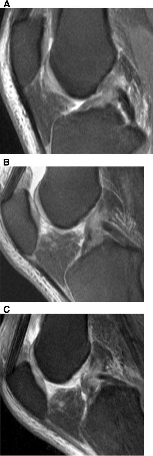
Arthroscopically proven partial ACL-rupture in a 53-year old patient after ski accident. (A) T2-weighted TSE FS sagittal image (2900/90) obtained at knee extension shows an irregularity of the midportion of the ACL. On T2-weighted TSE FS sagittal images (2900/90) at 30° of knee flexion (B) and at 55° of knee flexion (C) the partial continuity of the ACL bundles are better recognized than on MR images taken at knee extension (A).