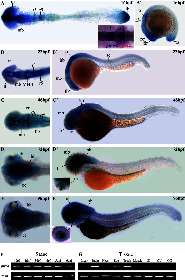
Fig. 1.
Spatial and temporal expression patterns of the zebrafish ptpro gene. Expressions of ptpro mRNAs were detected by antisense RNA whole-mount in situ hybridization (WISH). Images showing dorsal (a–d) or lateral views (a′–d′) of embryos collected at 16, 22, 48, 72, and 96 h post-fertilization (hpf). Anterior side is to the left and dorsal side is to the top. Small inserted image in (a) is from our double in situ staining to demonstrate the co-localization of the ptpro and ephA4a transcripts in the rhombomere 3 and 5. (f, g) Images of RT-PCR results for ptpro transcripts obtained from embryos at different developmental stages (f) or different adult tissues (g). Lower panels show RT-PCR results of α-actin for the controls. ce cerebellum, fb forebrain, hb hindbrain, MHB midbrain-hindbrain boundary, mb midbrain, re retina, r3/5 rhombomere 3/5, tb tailbud

