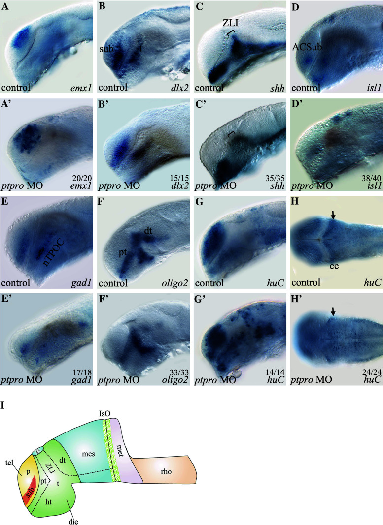
Fig. 3.
ptpro morphants exhibit defects in neuronal cell fate determination. (A-H’) Images of WMISH results from control (a–h) and ptpro MO-injected (a′–h′) embryos at 24 (a–g) and (a′–g′) and 72 hpf (h) and (h′). Each specific mRNA detected by WMISH is shown in the bottom right corner of each image. Lateral views with the anterior to the left and dorsal to the top in (a–g) and (a′–g′); dorsal views with the anterior to the left and right to the top in (h) and (h′). Arrow in (h′) indicates developing cerebellum. I Schematic drawing indicating the locations of dorsal thalamus (dt), diencephalon (die), epiphysis (e), hypothalamus (ht), isthmic organizer (IsO), metencephalon (met), pallial domain (p), prethalamus (pt), rhombencephalon (rho), subpallial domain (sub), telencephalon (tel), thalamus (t), zona limitans intrathalamica (ZLI). All embryos were injected with either 1 nl 1 % phenol red as injection control or 2 ng MO. The fraction of embryos displaying each phenotype is labeled on the corresponding image

