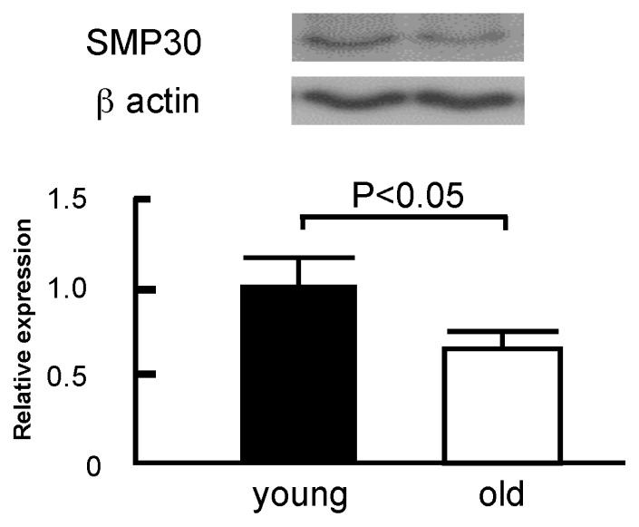
Figure A1.
SMP30 expression in myocardium of young and old mice. We measured SMP30 expression in myocardium of young (3 months old) and old (12 months old) mice (n = 5 each) by western blotting. Total protein was extracted from the snap-frozen left ventricle using Cell Lysis Buffer (Cell Signaling Technology, Inc., Beverly, MA, USA) with Protease Inhibitor Cocktail (BD Biosciences, San Jose, CA, USA) as previously reported [33]. Protein concentration was determined by protein assay (DC Protein Assay Kit, Bio-Rad Laboratories, Inc., Hercules, CA, USA). Equal amounts (20 μg) of the protein samples were separated by sodium dodecyl sulfate-polyacrylamide gel electrophoresis (SDS-PAGE, 5%–20%) and transferred onto polyvinylidene difluoride membranes (ATTO Co., Tokyo, Japan). The primary antibodies were anti-SMP30 (SHIMA Laboratories Co. Ltd., Tokyo, Japan) and mouse anti-β-actin (Santa Cruz Biotechnology Inc., California, CA, USA). The secondary antibodies were goat anti-rabbit IgG-horseradish peroxidase and goat anti-mouse IgG-horseradish peroxidase (Santa Cruz Biotechnology Inc., California, CA, USA). The signals from immunoreactive bands were visualized by an Amersham ECL system (Amersham Pharmacia Biotech UK Ltd., Buckinghamshire, UK) and quantified using densitometric analysis. SMP30 expression was lower in myocardium of old mice (12 months old) than young mice (3 months old). Data are expressed as the mean ± S.D.

