Figure 2.
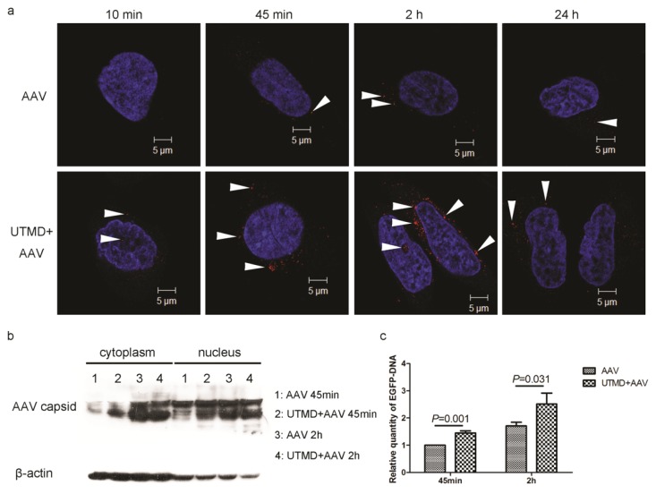
UTMD-mediated enhancement of AAV entry into HeLa cells. (a) Time-course of AAV uptake in HeLa cells with or without UTMD. AAV were added at an MOI of 2000 v.g./cell. Confocal immunofluorescence microscopy shows AAV (red, indicated by arrowheads) and nuclei (blue); (b) Quantities of AAV2 capsid protein in the cytoplasm or nuclei of AAV-transfected HeLa cells with and without UTMD at 45 min and 2 h post-infection; (c) Relative quantities of EGFP DNA following AAV transduction of HeLa cells with and without UTMD at 45 min and 2 h post-infection.

