
Figure 2.
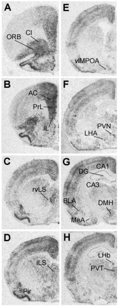
Representative autoradiographs of c-fos mRNA expression following novelty stress in a non-CVS exposed rat. The different levels from A to H represent anterior to posterior coronal brain sections. Abbreviations: AC, anterior cingulate cortex; BLA, basolateral nucleus of amygdala; CA1, CA1 region of hippocampus; CA3, CA3 region of hippocampus; Cl, claustrum; DG, dentate gyrus; DMH, dorsomedial hypothalamic nucleus; IL, infralimbic cortex; iLS, intermediate lateral septum; LHA, lateral hypothalamic area; LHb, lateral habenula; MeA, medial nucleus of amygdala; ORB, orbital cortex; Pir, piriform cortex; PrL, prelimbic cortex; PVN, paraventricular nucleus of hypothalamus; PVT, paraventricular nucleus of thalamus; rvLS, rostroventrolateral septum; vlMPOA, ventrolateral medial preoptic area.