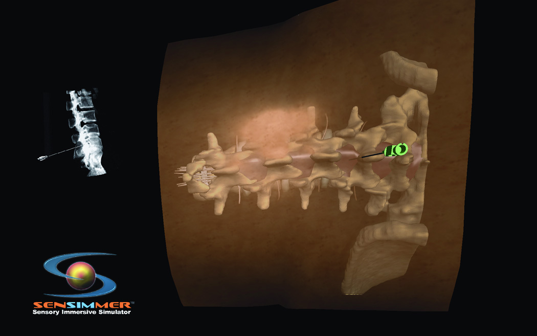
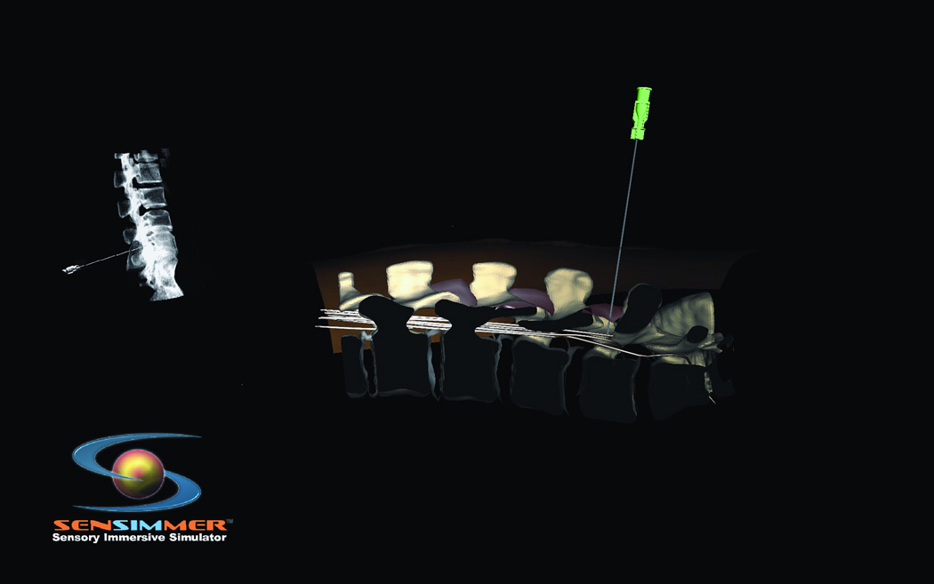
Figure 6.
(A) Virtual model for a patient’s back with the lumbar spine incorporated in the haptics of the model. The needle is aimed through the inter-laminar space. The location of the tip is seen on a virtual fluoroscopy screen. (B) At the end of the needle insertion, the operator perform a virtual sagital cut through the model to verify the trajectory and location of the tip of the needle.


