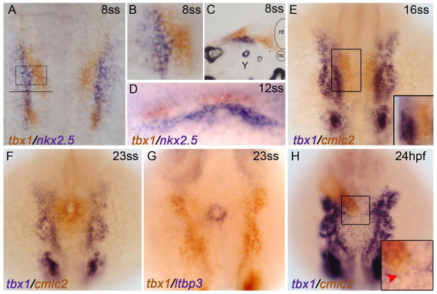
Figure 1. tbx1 transcripts fail to co-localize with cardiac cells during heart field or heart tube stages.
Whole-mount double in situ hybridization. (A–D) tbx1 (red) and nkx2.5 (blue) transcripts are non-overlapping during heart field stages (8 somite stage). (A) Flat mount, dorsal view, anterior up, 10X maginification. (B) 20X magnification of the ALPM (boxed region from A) (C) Transverse cryo-section, 20X magnification of the ALPM (location of section shown by the solid line in A) (D) Dorso-lateral view showing that tbx1-expressing cells lie dorsal to nkx2.5-expressing CPCs at the 12ss. (E,F) tbx1+ cells are lateral to medially migrating cmlc2+ cardiomyocytes at 16ss and 23ss. (G) tbx1+ cells are lateral to ltbp3+ SHF progenitors at 20.5hpf/23ss. Dorsal view, anterior up. (H) At 24hpf, the linear heart tube lies dorsal to the tbx1 expression domain. Dorsal view, anterior up. 10X magnification. (inset) 20X magnification of boxed region from F. tbx1 is not expressed at the end of the linear heart tube where SHF progenitors reside (red arrowhead).

