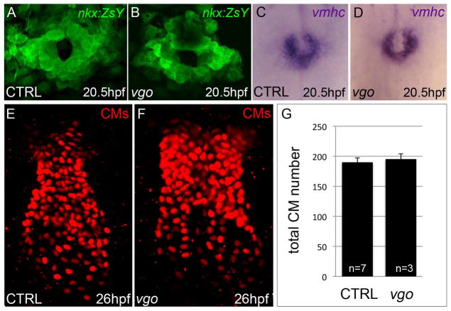
Figure 3. Cardiac progenitors within the heart forming region are specified in tbx1 mutants and differentiate appropriately into ventricular cardiomyocytes.
(A,B) Flattened confocal images of Tg(nkx2.5::ZsYellow) control (n=37; A) and vgo (n=14; B) embryos at 20.5hpf/23somite-stage (ss). (C,D) Whole-mount in situ hybridization of vmhc at 20.5hpf/23ss in both control (n=16; C) and vgo (n=16; D) embryos. (E,F) Flattened confocal images following immunofluorescence at 26hpf to visualize DsRed+ nuclei in control (E) and vgo (F) embryos. Dorsal view, Anterior up in all images. (G) Graph depicting the average number of CMs at 26hpf in control (n=7) and vgo (n=3) embryos.

