
Figure 5. SHF progenitor cells fail to proliferate in the absence of Tbx1.
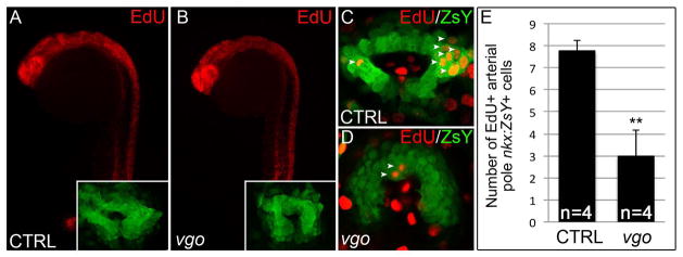
(A–D) Click-iT EdU labeling in Tg(nkx2.5:ZsYellow) vgo and control siblings. (A–B) Flourescent microscopy images of EdU+ cells (red) in control (A) and vgo (B) embryos. 10X maginificaition, anterior up, dorsal right. Insets show flattened confocal images of ZsY+ cells at the arterial pole (C–D) Composite of two confocal sections showing EdU+ cells (red) within the ZsYellow+ (green) SHF (white arrowheads). (E) Graph depicting the total number of EdU+ cells in the entire confocal stack in control (n=4) and vgo (n=4) embryos.