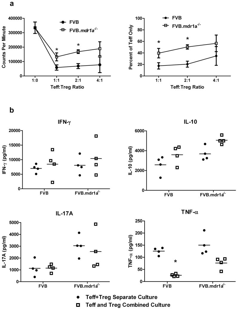
Figure 2.
FVB.mdr1a−/− CD4+CD25+ Treg cells suppress CD4+CD25− Teff proliferation in vitro, but fail to suppress TNF-α secretion. (a) MACS isolated FVB CD4+CD25− (Teff) and CD4+CD25+ (Treg) cells from either FVB or FVB.mdr1a−/− were cultured either separately or cultured together for 96 hours in the presence of anti-CD3 and anti-CD28. 3H-Thymidine was added for the final 24 hours of culture. Assay was done at 1:1, 2:1, and 4:1 Teff:Treg ratio as indicated. (b) Supernatants from a were collected after 72 hours and prior to the addition 3H-Thymidine. Cytokine concentrations were determined using multiplex kits. Data are representative of 3 separate experiments, with 3–4 male mice, 6–8 weeks of age in each group per experiment. * P ≤ 0.05; ** P ≤ 0.01; *** P ≤ 0.001. Mean + standard deviation (a) or standard error of the mean (b) shown.

