Figure 3.
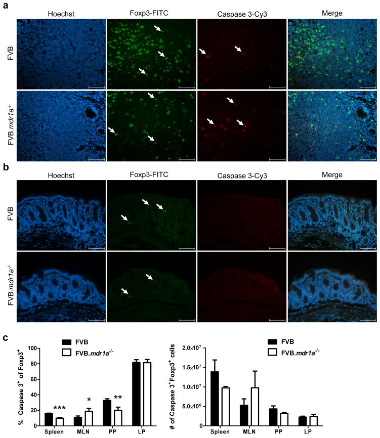
Intestinal FVB.mdr1a−/− Foxp3+ cells do not have increased levels of apoptosis. (a) Sections of Peyer’s patches and (b) distal colon were stained for Foxp3 and cleaved caspase-3. Positive cells for stains are indicated by arrows in their respective panels. Images were captured at 40× magnification, and scale bars = 20 μm. (c) Cells were isolated from spleen, MLN, PP, and intestinal LP and stimulated for 18 hours with 10 μg/ml anti-CD95 to induce apoptosis. Cells were then stained for CD4, Foxp3 and cleaved caspase-3. Lymphocytes were gated based on forward scatter/side scatter. Cells were pre-gated on CD4+ cells prior to analysis of Foxp3 and cleaved caspase 3 expression. Note that the high number of apoptotic cells in the LP may be a result of the extensive enzymatic digesting necessary to isolate these cells. Data are shown from one experiment, but are representative of 2 separate experiments, with 3–4 male mice, 6–8 weeks of age in each group per experiment. * P ≤ 0.05; ** P ≤ 0.01; *** P ≤ 0.001. Mean + standard deviation shown.

