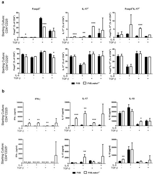
Figure 6.
FVB.mdr1a−/− CD4+CD25− cells do not become Foxp3+ cells upon treatment with TGF-β and are more likely to become Foxp3−IL-17+ cells. (a) MACS isolated CD4+CD25− (upper panels) and CD4+CD25+ (lower panels) cells were cultured for 72 hours with CD3 and CD28 stimulation in the presence of 25 ng/ml IL-6 alone, 5 ng/ml TGF-β alone, or IL-6 and TGF-β together. Cells were rested for 24 hours, then subsequently stained for CD4, Foxp3, and IL-17. (b) Supernatants from a were collected after 72 hours of culture and analyzed for IL-10, IL-17, and IFN-γ secretion by ELISA. It is important to note that freshly isolated splenic CD4+Foxp3− (and CD4+Foxp3+) cells from FVB and FVB.mdr1a−/− express similar levels of the activation markers CD44, CD62L, and CD69, indicating that the activation states of these cells is the same (data not shown). Results are representative of 2 independent experiments, with 3–4 male mice, 6–8 weeks of age in each group per experiment. N.D. = not detected (below detectable level of assay). * P ≤ 0.05; **, P ≤ 0.01; ***, P ≤ 0.001. Mean of 3–4 mice (analyzed separately) + standard deviation shown.

