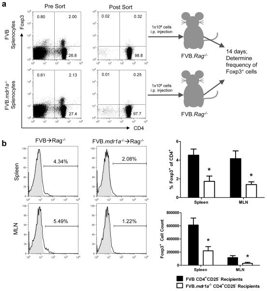
Figure 7.
FVB.mdr1a−/− CD4+CD25− cells show impaired Foxp3+ iTreg generation in vivo. (a) An schematic of the adoptive transfer experiment. Splenocytes from FVB and FVB.mdr1a−/− mice were stained for CD4 and CD25, and subsequently isolated via FACS cell sorting. Representative pre-sort (left panels) and post-sort (right panels) are shown for both FVB and FVB.mdr1a−/−. 1×10⁶ CD4+CD25− cells were then injected i.p. into FVB.Rag2−/− recipients. After 14 days, the spleen, MLN and intestinal lamina propria was harvested from the recipients. (b) Foxp3+ expression on the gated CD4+ population of spleen and MLN of FVB.Rag2−/− recipients 14 days after adoptive transfer. * P ≤ 0.01. Mean of 6 recipient mice per group + standard deviation shown.

