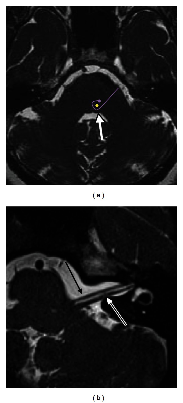
Figure 1.

Normal facial nerve on MRI. (a) Axial CISS image at the level of the pons demonstrates the facial colliculus (arrow) seen as a small bump along the anterior wall of the fourth ventricle. This is formed by the motor tracts of the facial nerve (purple curved line) coursing around the abducens nucleus (yellow dot). (b) Axial CISS sequence of the left CPA and IAC demonstrates the normal cisternal and intracanalicular segments of the left CN VII (solid arrow), anterior to CN VIII (double lined arrow).
