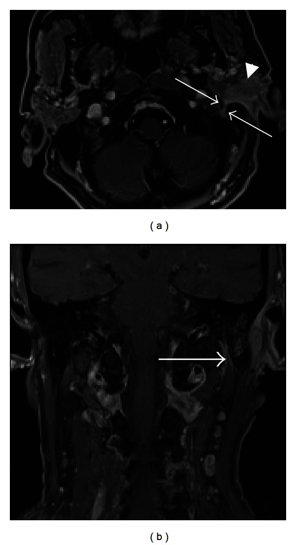
Figure 9.

Perineural tumor spread along CN VII. Axial T1 (a) and coronal (b) postcontrast fat-saturated images demonstrate an enlarged, enhancing left facial nerve in the stylomastoid foramen and mastoid segment of the facial nerve canal (arrows), indicating perineural tumor spread from invasive squamous cell carcinoma (arrowhead) of the left external auditory canal.
