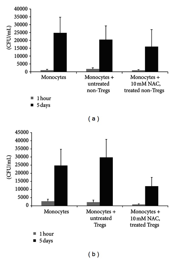
Figure 7.

Determination of intracellular viability of H37Rv in cocultures of H37Rv-infected monocytes and CD4+ T cells (non-Tregs and Tregs). Tregs and non-Tregs were isolated from PBMCs derived from healthy subjects using midi-MACS LD columns and mini-MACS MS columns from Miltenyi Biotech. Adherent monocytes were infected with processed H37Rv at a multiplicity of infection of 1 : 1 and incubated for 2 hours for phagocytosis. Unphagocytosed mycobacteria were removed by washing the infected monocytes three times with sterile PBS. Infected monocytes were cultured in RPMI containing 5% AB serum in presence and absence of autologous non-Tregs (a) and Tregs cells (b). Prior to co-incubation with infected monocytes, autologous CD4+ T cells (both Tregs and non-Tregs) were incubated overnight with NAC (10 mM), washed with PBS, re-suspended in fresh RPMI containing AB serum (without any additives), and then added to the infected monocytes (monocyte: T cell ratio was adjusted to 1 : 1). Infected monocyte-CD4+ T cell cocultures were terminated at 1 hour and 5 days after infection to determine the intracellular survival of H37Rv. Infected monocytes cultured in the absence of T cells served as negative controls. Results were analyzed using one-way ANOVA with Dunnett's multiple comparisons test, comparing all T-cell co-culture categories to the monocyte only control. Our results did not achieve statistical significance.
