Figure 1.

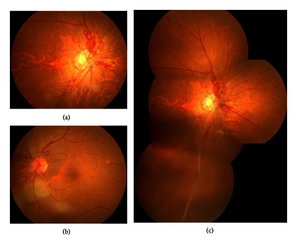
Fundus photographs showing a bilateral prepapillary vascular loop (a, b) and persistent hyperplastic primary vitreous in the right eye (c).

Fundus photographs showing a bilateral prepapillary vascular loop (a, b) and persistent hyperplastic primary vitreous in the right eye (c).