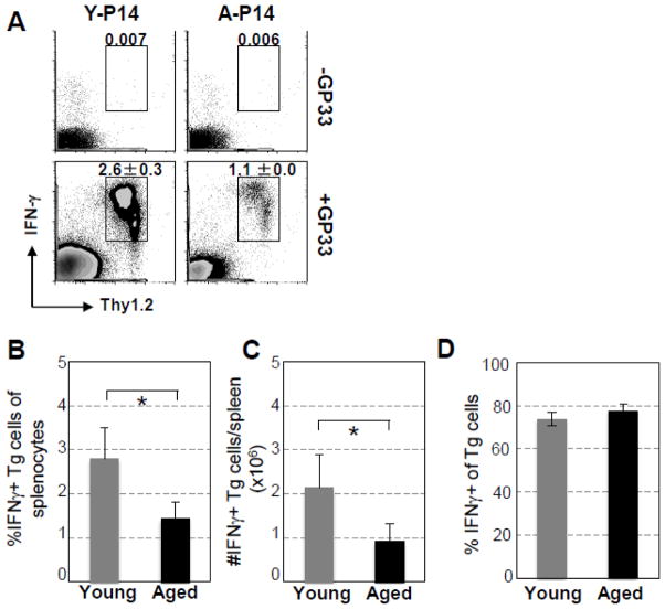
Fig. 5.
Decreased clonal expansion, but not producing IFN-γ function, of Tg CD8 T cells of aged P14 mice transferred to young recipient mice challenged with LCMV. Similar to Fig. 4, on Day 7 after infection, splenocytes were harvested and stimulated ex vivo with GP33-41 peptide for 4 h. IFN-γ production of transferred P14 cells was examined by surface and intracellular IFN-γ staining, respectively. (A). A representative experiment shows the percentages specific Tg CD8 T cells of splenocytes secreting IFN-γ. (B). Percentages, (C) absolute numbers of IFN-γ secreting Tg CD8 T cells in spleens, and (D) percentages of Tg CD8 T cells that secrete IFN-γ of transferred Tg CD8 T cells of 15 mice pooled from 4 independent experiments. * p < 0.05.

