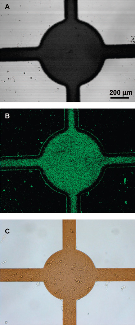
Figure 4.
Selective adsorption of proteins and cells onto ITO electrodes after electrical “switching” of surface properties. (A) ITO electrode (dark region) fabricated on glass (100× magnification). (B) Selective adsorption of collagen–FITC to the activated ITO electrode. Note lack of protein deposition to glass regions that remain nonfouling due to the presence of PEG silane. (C) Preferential attachment of hepatic stellate cells to the activated electrode regions and not to glass domains that retain PEG silane layer.

