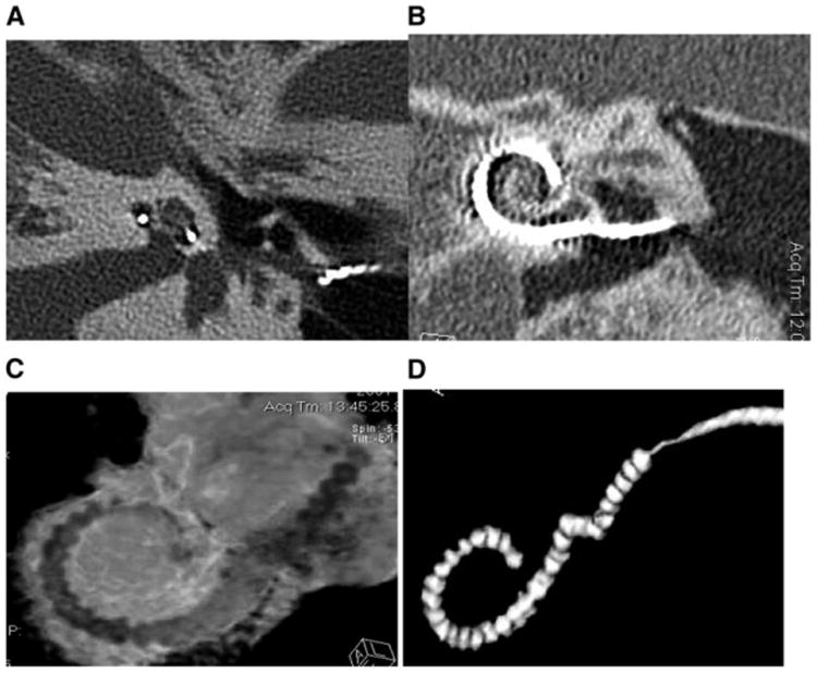
Figure 4.
The images show (A) a midmodiolar scan of an implanted right ear from which cochlear radii and height can be measured; (B) a 2-D reformatted image to show a coronal view of the implant in vivo; (C) a 3-D reconstruction of the implant within the cochlear canal (Supplemental to the online version of this article is a version of this figure in which electrodes are shown in green, the intracochlear tissues in red, and the cochlear capsule transparent with canal walls in white); (D) shows a 3-D of the array extracted digitally with metal artifact reduction applied to provide better definition of the individual electrode positions. Note in A, B, and C the shift in position of the trajectory of the array from the outer wall of the cochlear canal to the modiolar wall in the upper basal turn.

