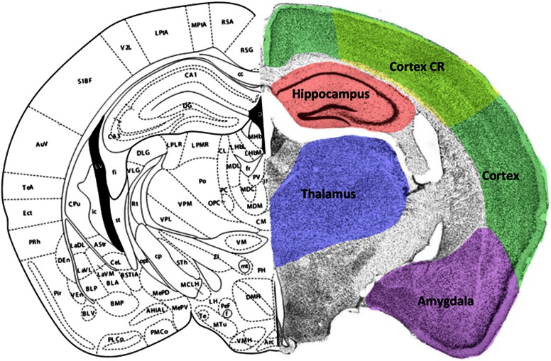
Figure 1.
Coronal brain section through the dorsal hippocampus illustrating regions of interest (ROIs) examined in this study. Left hemisphere taken from Franklin and Paxinos23 at approximately 2.0 mm caudal to Bregma. Right hemisphere illustrates a cresyl violet-processed section with ROI outlined in color. The color reproduction of this figure is available on the JCBFM journal online. CR, contusion region.

