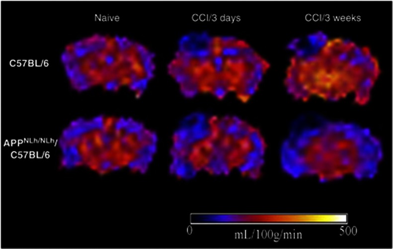
Figure 2.
Representative cerebral blood flow (CBF) maps illustrated on coronal views of arterial spin-labeling MR images taken at the level of the dorsal hippocampus from naive and injured C57 and APPNLh/NLh mice. CBF levels in each region of interest defined in Materials and Methods were quantified and are illustrated in Figure 3. CCI, controlled cortical impact.

