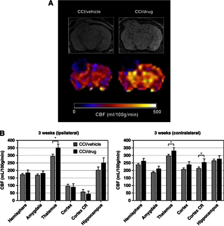
Figure 4.
(A) Representative magnetic resonance image (A, top row) cerebral blood flow (CBF; A, bottom row) maps illustrated in coronal views at the level of the dorsal hippocampus from APPNLh/NLh mice after controlled cortical impact (CCI) injury and 3 weeks treatment with either vehicle or 3 mg/kg simvastatin. (B) Region of interest CBF values from the two experimental groups with CCI/vehicle (gray bars) and CCI/drug (black bars) treatment. *CCI/drug>CCI/vehicle, P<0.05. CR, contusion region.

