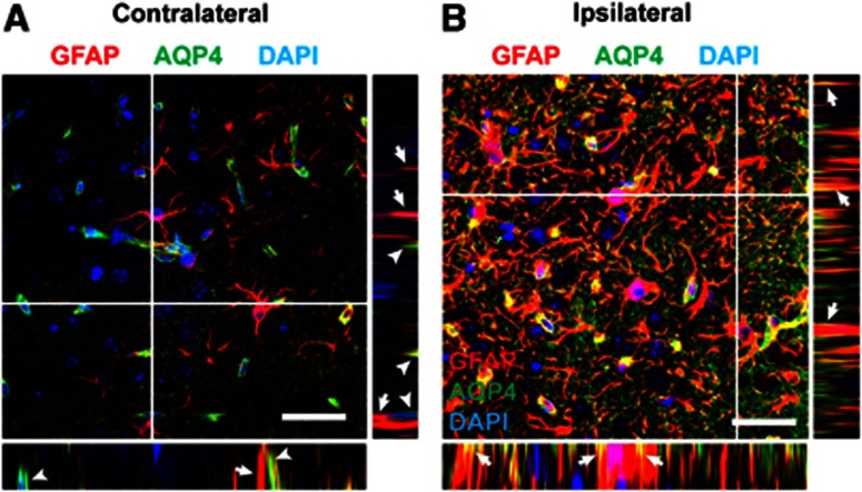
Figure 6.
Post-traumatic redistribution of AQP4 localization. Localization of AQP4 was assessed by high-power confocal microscopy 14 days after moderate TBI. (A) On the contralateral side, AQP4 immunoreactivity is confined to perivascular endfeet (arrowheads) and generally does not co-localize with large glial fibrillary acidic protein (GFAP)-positive astrocytic processes (arrows). Insets depict XZ and YZ projections at planes indicated by white lines. (B) After moderate traumatic brain injury (TBI), a large proportion of AQP4 immunoreactivity has shifted to the membrane of GFAP-positive reactive astrocyte soma and coarse processes (arrows). AQP4 immoreactivity is also evident diffusely surrounding GFAP-positive processes, presumably corresponding to fine astrocytic processes. Insets depict XZ and YZ projections at planes indicated by white lines. Scale bars: 25 μm.

