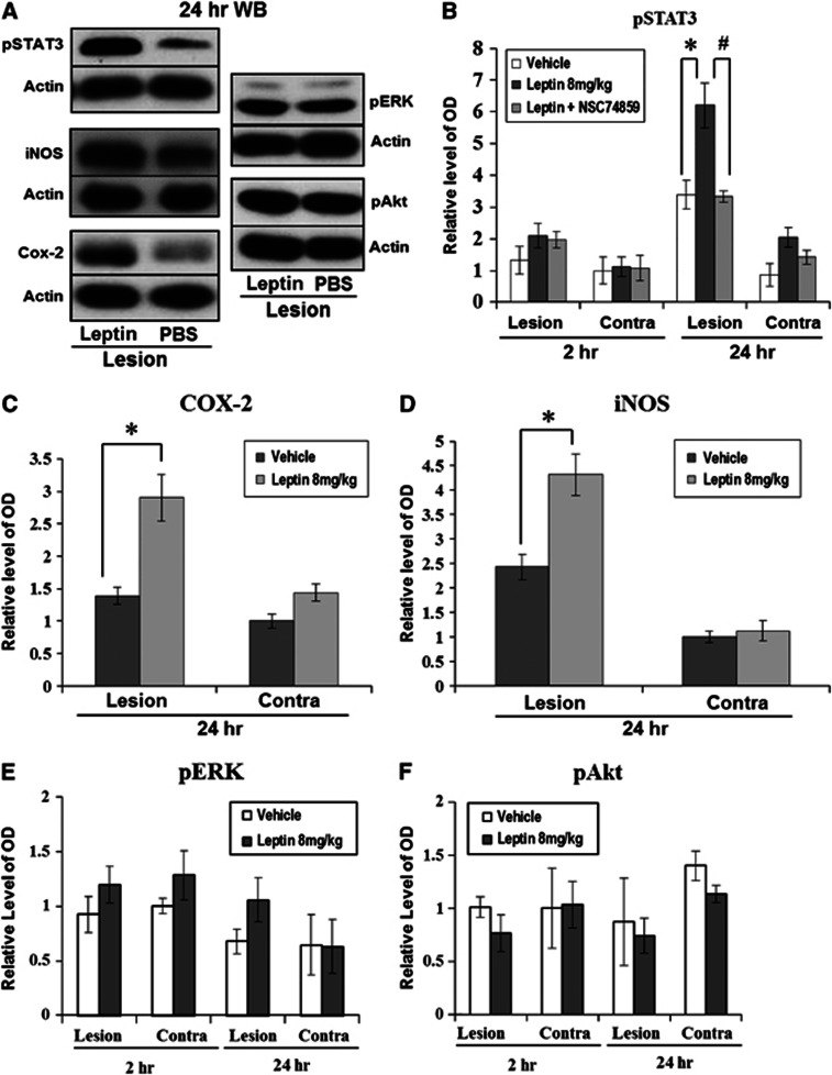
Figure 5.
Western blot analyses for signal transduction and activator of transcription 3 (STAT3), extracellular signal-regulated kinase (ERK), Akt, and cyclooxygenase-2 (COX-2). (A) At 24 hours after intracerebral hemorrhage (ICH), the density of pSTAT3 and COX-2 in the leptin-injected group was higher than in the control group (n=4 each). (B) In quantitative analyses, pSTAT3 was minimally detected at 2 hours after ICH, but 24 hours later, a 1.8-fold increase of pSTAT3 density was detected in the leptin-injected group (*P<0.05). After the injection of NSC74859, a specific inhibitor of pSTAT3, the pSTAT3 density in the leptin-injected group was significantly decreased, nearly to the level of the control group (#P<0.05). (C) The density of COX-2 in the hemorrhagic hemisphere showed a 2.1-fold increase in the leptin-injected group compared with the phosphate-buffered saline (PBS)-injected group. (D) The density of inducible nitric oxide synthase (iNOS) in the hemorrhagic hemispheres showed a 1.8-fold increase in the leptin-injected group compared with the PBS-injected group. (E, F) The densities of pERK and pAkt were not different between the leptin-injected and control groups.

