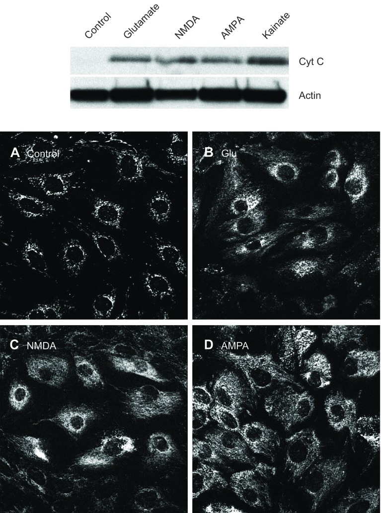
Fig. 8.
Detection of cytochrome c. Confluent quiescent CMVEC were exposed to glutamate (1 mM), NMDA (0.1 mM), AMPA (0.1 mM), or kainate (0.1 mM) for 3 h. Cytochrome c in control and treated cells was detected by immunoblotting in mitochondria-free cytosolic fraction (top) and by immunofluorescence in paraformaldehyde-fixed cells (A–D).

