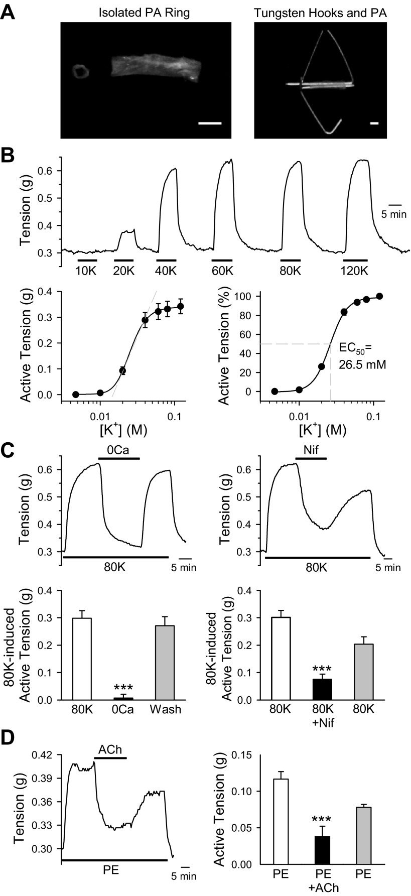
Fig. 1.
Ca2+ influx through voltage-dependent Ca2+ channels (VDCC) plays an important role in pulmonary vasoconstriction. A: image shows intrapulmonary artery isolated from mice (left). Two tungsten hooks were carefully passed through the lumen of pulmonary artery (PA) rings (right) to measure isometric tension. B: PA rings were exposed to modified Krebs solution (MKS) containing extracellular K+ concentrations of 10, 20, 40, 60, 80, and 120 mM. Representative tension tracings are shown at top. Summarized absolute active tension and active tension normalized to the maximum tension generated by 120 mM K+ are shown at bottom. C: mouse PA were treated with Ca2+ free (left) or 300 nM nifedipine (Nif; right) on the 80 mM K+ (80K)-induced contraction. After peak contraction was achieved, arteries were exposed to either Ca2+ free (0Ca) or 300 nM Nif to attenuate Ca2+ influx. Summarized data showed active tension induced by 80K before (80K), during (0Ca), and after (Wash) exposure to 0Ca (bottom left) or Nif-containing solution (bottom right). D: representative tension record showing phenylephrine (PE; 100 nM)-induced active tension before, during, and after application of acetylcholine (ACh; 10 μM). After peak contraction with PE was achieved, arteries were exposed to ACh (left). Summarized data showed absolute tension after application of PE, ACh, and washout (right). Data are expressed as means ± SE. Rings were isolated from 5–7 mice. ***P < 0.001, statistical difference from the control value.

