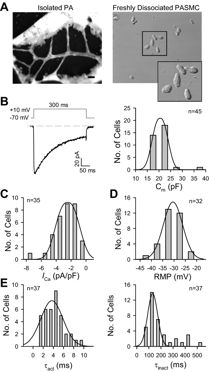
Fig. 2.
Passive membrane properties of freshly dissociated mouse PA smooth muscle cells (PASMC). A: images show the isolated pulmonary artery from the left lobe in a 7- to 8-wk-old male mouse (left) and single PASMC dissociated from the 2nd-3rd orders of intrapulmonary arterial branches isolated from mice (right). Scale bar = 800 μm. B: representative currents were evoked by a depolarizing pulse to +10 mV from a −70-mV holding potential (left). Histogram shows the distribution of membrane capacitance (Cm, n = 45 cells; right). C: peak currents (or current density) were determined at +10 mV, normalized to cell capacitance (n = 35). D: averaged resting membrane potential (RMP; n = 32) in freshly dissociated mouse PASMC. E: activation and inactivation time constants, τact (left) and τinact (right), were measured by fitting a monoexponential curve to the rising phase (n = 37) and decaying phase (n = 37) of each type of current. Cells were isolated from 30–42 mice.

