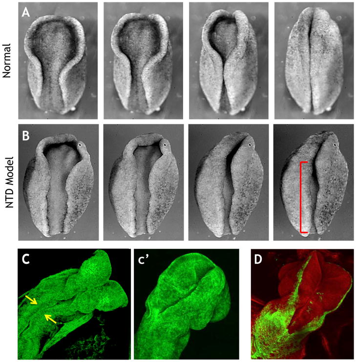
Figure 2.
A. Still frames from a time lapse movie showing neural tube closure in an amphibian embryo (rostral = up). B. Disruption of PCP signaling results in disrupted neural tube closure (red bracket). C. Still frame from a movie of mouse neural tube closure; arrows indicate initial meeting of neural folds (47). c′. Neural tube closure has progressed in a later time point. D. Genetically-inducible fluorescent reporters allow visualization of specific tissues (green), in this case the ectoderm that borders the neural tissue.

