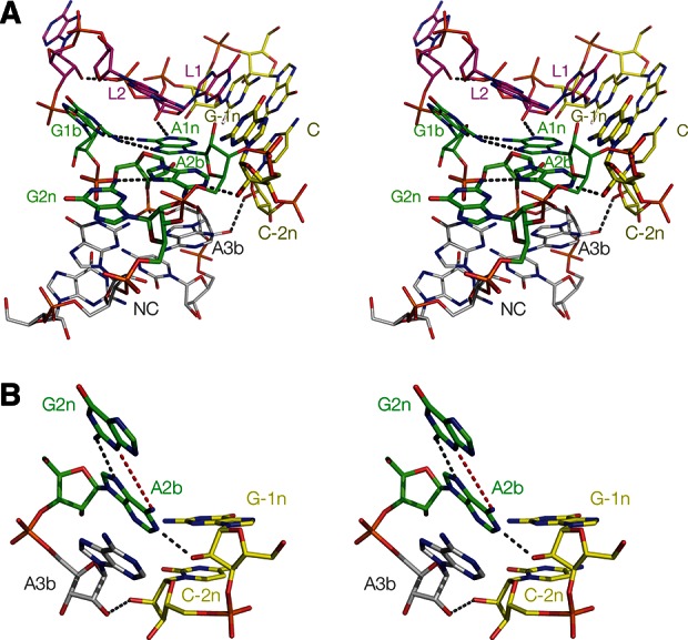
FIGURE 3.

Parallel-eye stereo images of Kt-7 in the H. marismortui 50S ribosomal subunit. (A) The whole k-turn viewed from the side of the nonbulged strand. Key hydrogen bonds are shown as gray broken lines. (B) Nucleotides participating in the key A-minor interactions in the core of the k-turn. Note that G−1n donates its O2′ proton to N1 of A2b, i.e., this is an N1-class k-turn. As a consequence, the distance between A2b N6 and G2n N3 is too long for hydrogen bonding, at 4.3 Å (shown in red). There is an additional hydrogen bond between the O2′ groups of C−2n and A3b that is never observed in the N3 structures.
