
Figure 2.
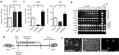
Activation of the PI3K-Akt pathway enhances iPSC generation. (a) Counts of AP+ colonies formed by transduction of OKSM combined with the dominant-negative form of Pten (CS-Pten) or activated form of Akt (myr-Akt) into MEFs. Wild-type MEFs (1 × 10⁵) were transduced with OKSM (control, left), OKSM+CS-Pten (middle), or OKSM+myr-Akt (right). Cells (5,000) were transferred onto SNL feeders on day 4 after transduction and cultured in ESC medium. AP+ colonies were counted on day 14 after transduction. Data are the mean ± SD (n = 3), *P < 0.05 versus control. (b) Experimental scheme for iPSC generation. MEFs (1 × 10⁵) were infected with retroviruses carrying OKSM on day 0. Cells (5,000) were transferred onto SNL feeders on day 4 after transduction and cultured in ESC medium containing 100 nmol/l bpV(HOpic). Colonies were collected based on ESC-like morphology on day 14 after transduction. The Pten inhibitor bpV(HOpic) was added from day –1 to 10. (c) Counts of AP+ colonies formed by transduction of OKSM or OKS into MEFs in the presence of LY294002 or bpV(HOpic). A total of 5,000 (OKSM) or 50,000 (OKS) retrovirally transduced MEFs were transferred onto SNL feeders on day 4 after transduction and then cultured in the presence of 100 nmol/l bpV(HOpic) or 5 μmol/l LY294002 for 10 (OKSM) or 28 days (OKS). AP+ colonies were counted on day 10 (OKSM, left panel) and day 28 (OKS, right panel). AP+ colonies generated without drugs were used as controls. Data are the mean ± SD (n = 3), *P < 0.05 versus controls. (d) Characteristics of bpV-iPSCs in vitro. The expression of ESC marker genes in bpV-iPSCs (OKSM) was examined by RT PCR (bpV-iPSC clones 1–8). Mouse β-actin was used as a loading control. The RT (−) control is shown at the bottom. (e) Representative images of bpV-iPSC colonies (left panel), AP staining (middle panel), and SSEA1 staining (right panel). Scale bars = 100 μm. ESC, embryonic stem cell; iPSC, induced pluripotent stem cell; MEF, mouse embryonic fibroblast; RT, reverse transcription.