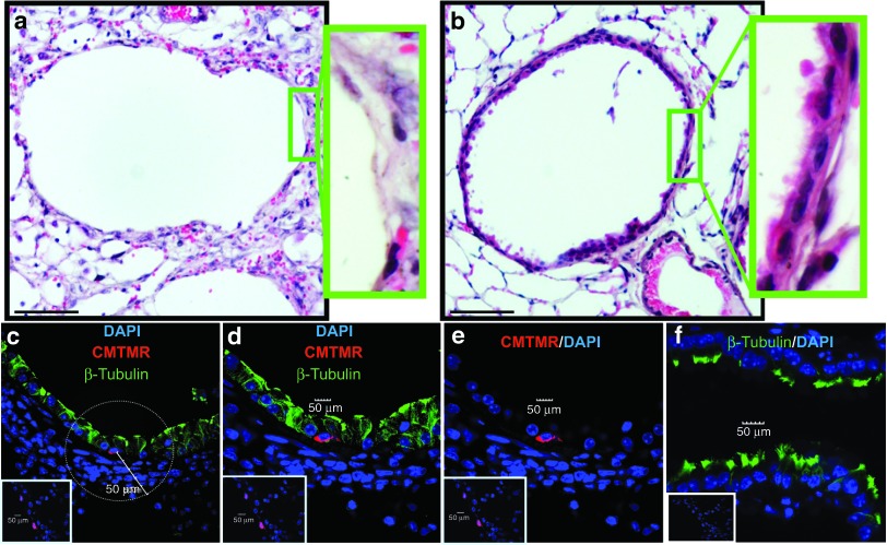
Figure 3.
CCSP+ BMC preserve the epithelium in large airways. (a) Mice treated with CCSP− BMC have a poorly preserved epithelium compared with (b) mice treated with CCSP+ BMC. Green insets: higher magnification image of boxed region. (c–e) Confocal microscopic analysis of the ciliated cell marker β-tubulin IV (green fluorescence) showed that donor cells (CMTMR+; red fluorescence) do not express β-tubulin but are preferentially surrounded by host β-tubulin+ cells (c1 and c2) in the CCSP+ BMC group. (f) The host β-tubulin+ cells exhibited reduced numbers of organized cilia on the cell surface which make them fluorescent not only in the apical region but also in the cytoplasm in contrast to ciliated cells from normal lung that only showed β-tubulin expression in the apical cilia. Insets are representative isotype staining controls. Scale bar represents 10 µm. n = 4 mice per group. Samples from mice administered with donor cells after 10 days of ganciclovir. BMC, bone marrow cells; CCSP, Clara cell secretory protein.

