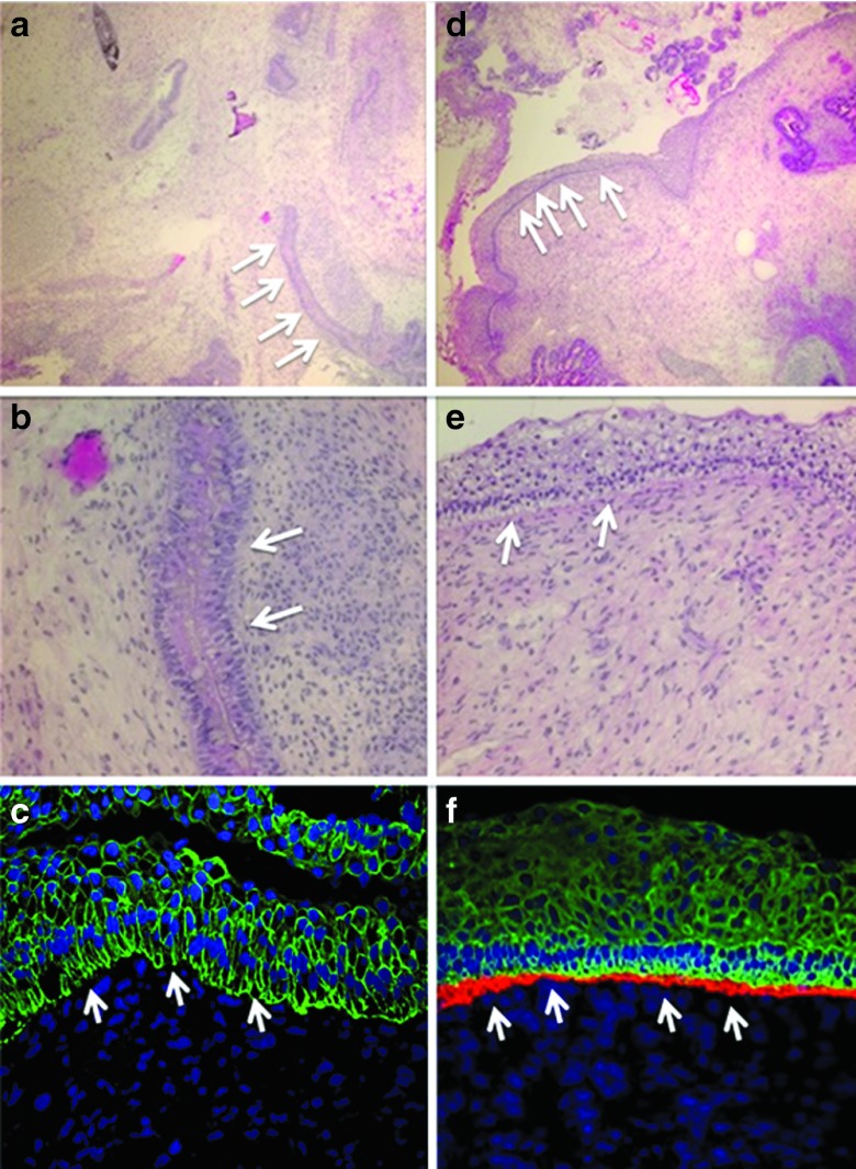
Figure 7.
iPSC teratoma and rescued type VII collagen contribution to the DEJ using gene-edited cells. iPSC-derived teratoma analysis. Homozygous 1837 C>T RDEB and TALEN gene-corrected fibroblasts were reprogrammed into iPSCs for teratoma differentiation assay. RDEB mutant cells: (a,b) iPSC light microscopy and (c) immunofluorescence. RDEB TALEN-corrected cells: (d,e) iPSC light microscopy and (f) immunofluorescence. (a,b,d,e) Hematoxylin and esosin stained light microscopy images are at 10× and 20× magnification, respectively and showed epidermal skin-like structures (white arrows). (c,f) Teratoma immunofluorescence shows the DEJ indicated with white arrows. Immunofluorescence markers were as follows: Blue, DAPI nuclear stain; green, cytokeratin 5; red, type VII collagen. Images are representative images from at least three animals. Antibody staining was performed from a single master mix on the same day using identical microscopy settings.

