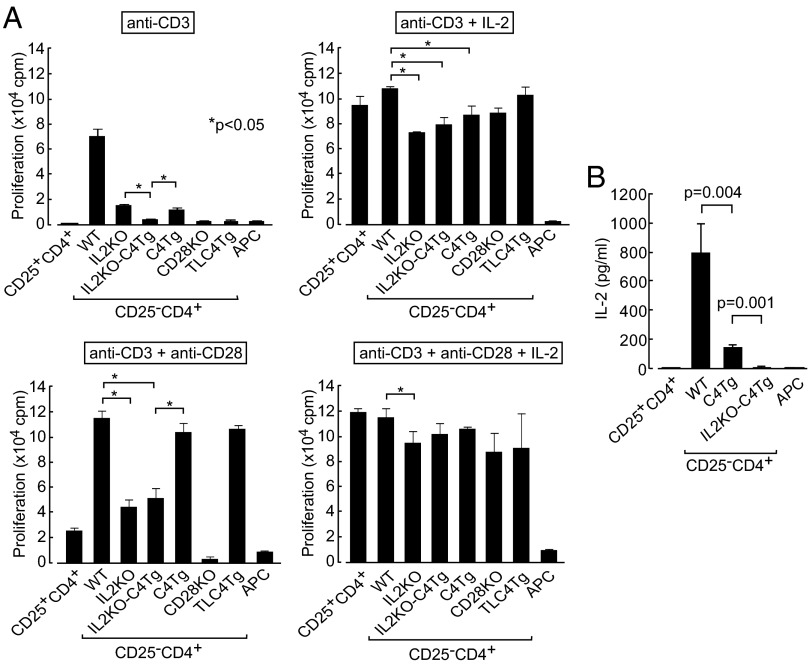
Fig. 3.
Proliferation of IL2KO-C4Tg CD4+ T cells. (A) Proliferation measured by 3H-thymidine incorporation of CD25+ or CD25−CD4+ lymph node T cells from designated groups of mice when stimulated for 3 d in the presence of APCs and indicated reagents. Significant difference were analyzed with post hoc comparison in ANOVA among IL2KO, IL2KO-C4Tg, and C4Tg Tn cells under anti-CD3 stimulation and among WT, IL2KO, IL2KO-C4Tg, and C4Tg Tn cells in the other conditions. cpm, count per minute. (B) IL-2 concentration assessed by ELISA in culture supernatant after 3-d incubation in the presence of APCs and anti-CD3. Representative results (mean ± SD of triplicates) from three independent experiments are shown. TLC4Tg mice used were deficient in endogenous CTLA-4 expression by mating with CTLA-4−/− mice.

