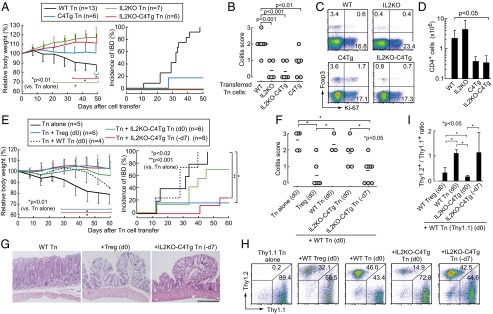
Fig. 6.

Suppression of colitis in RAG2−/− mice by IL2KO-C4Tg CD4+ T cells. (A) Body weight change (mean ± SDs) and incidence of IBD in RAG2−/− mice transferred with 1 × 10⁵ CD45RBhighCD25−CD4+ Tn cells from indicated groups of mice. (B) Severity of colitis based on histological evaluation in each group of recipient mice 50 d after cell transfer. Circles represent individual mice. Horizontal bars indicate the means. (C) Intracellular Ki-67 and Foxp3 expression of transferred CD4+ T cells in mesenteric lymph nodes of recipient mice 50 d after cell transfer. (D) Numbers (mean ± SDs) of CD4+ T cells in mesenteric lymph nodes in the groups of mice shown in B. (E–I) Indicated numbers of RAG2−/− mice received 1 × 10⁵ Thy1.1+ CD45RBhighCD4+ Tn cells on day 0 along with the same numbers of WT CD25+CD4+ Treg cells or CD45RBhighCD4+ Tn cells from designated groups of mice. Pretransfer of indicated Tn cell populations was performed on day −7 (−d7) in some mice as depicted. (E) Body weight change (mean ± SD) and incidence of IBD, (F) histological score of colitis, and (G) representative histology (H&E staining) of the colon from each group of recipient mice. (Scale bar, 20 μm.) (H and I) Thy1.1/Thy1.2 staining (H) and the ratio in CD4+ T cells (I) in mesenteric lymph nodes of recipient mice (shown in F) 60 d after cell transfer.
