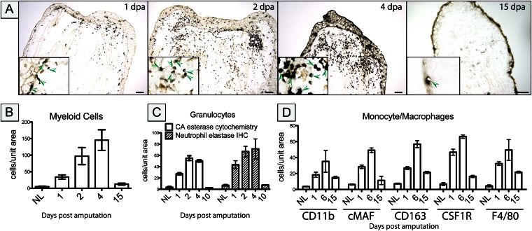
Fig. 2.
Myeloid cells accumulate in the regenerating limb blastema. (A) α-Naphthyl acetate (NSE) enzyme cytochemistry was used to detect myeloid cells at different time points after amputation. Cell numbers were determined by counting NSE-positive cells to detect myeloid cells (B), CA esterase cytochemistry to detect granulocytes (C), and immunocytochemistry to detect monocytes/macrophages (D), using Fiji image analysis software on at least three separate experiments. Cell numbers are expressed as the number of cells counted per unit area as the mean ± SEM. Representative sections are shown in Figs. S2 and S3. (Scale bars, 100 μm.)

