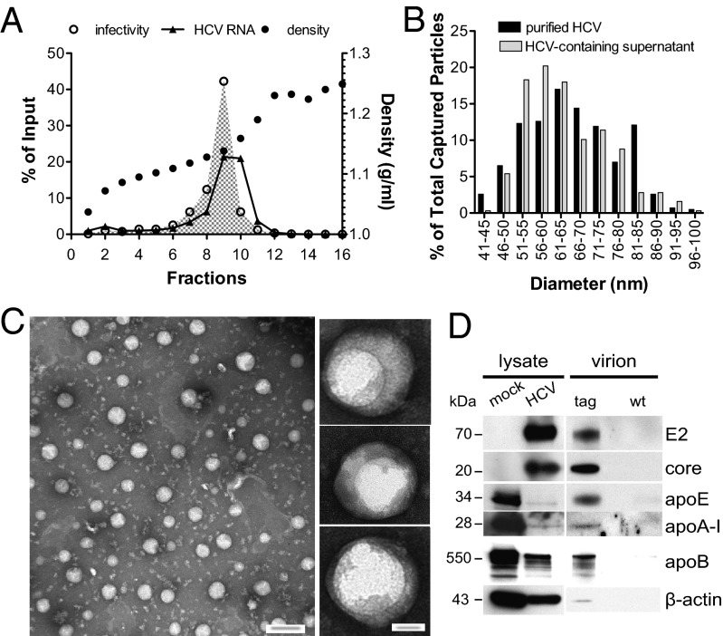
Fig. 6.
Biophysical and ultrastructural characterization of purified HCV. (A) High-titer, heparin-purified tag-HCV virions were fractionated according to density on a 10–40% (wt/vol) iodixanol gradient. For each fraction, viral RNA, infectivity, and density were determined and expressed as percent of input. (B) Size distribution of highly infectious tag-HCV particles in fraction 9 (1.13 g/mL) of the buoyant density gradient (black bars; n = 430; mean = 67 nm, SD = 12 nm) is compared with unpurified HCV-containing supernatant (gray bars; n = 317; mean = 64 nm, SD = 11 nm). Data are expressed as percent of total captured particles. (C) Left: Low-magnification TEM image of tag-HCV virions from fraction 9 captured on 2% (vol/vol) Ni-NTA affinity grids. (Scale bar: 100 nm.) Right: Close up views of structures observed in purified preparations with high specific infectivity (Scale bar: 20 nm.) (D) Immunoblot for the indicated proteins in mock- or HCV RNA–electroporated Huh-7.5.1 cells, tag-, and WT-HCV extracellular virions that were heparin- and iodixanol-purified, precipitated with Dynabeads and eluted with 0.9 M imidazole.

