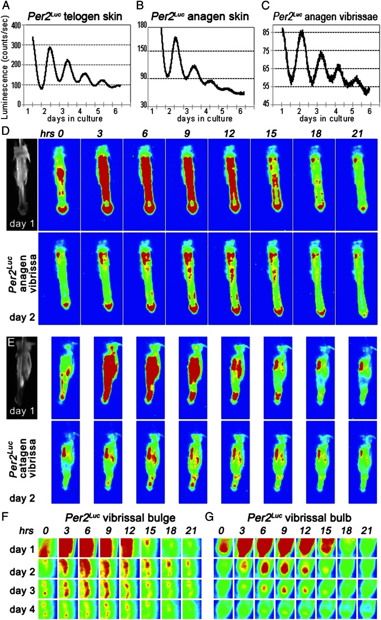
Fig. 1.
Per2Luc reveals peripheral circadian rhythms in skin and anagen hair follicles. (A and C) In cultured Per2Luc mouse skin with either telogen (A) or anagen (B) hair follicles, luminescence levels change with circadian periodicity for at least 3–4 d. (C) This circadian periodicity is also displayed by cultured Per2Luc vibrissae. (D–G) Time-lapse photography of individually cultured Per2Luc vibrissae shows that circadian cycles of luminescence localize to several follicular areas, most prominently to follicular bulge (F) and bulb (G). To aid visualization, originally black-and-white images of luminescence levels were converted into heat maps, so that areas of highest luminescence appear red, and areas with no luminescence appear blue, with blue–green–yellow–red gradient in between. Sequential snapshots shown here are 3 h apart. Microdissected vibrissae at time point zero are shown for D and E. Also see Fig. S1.

