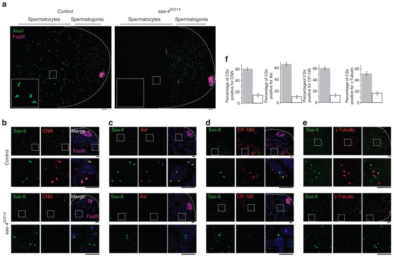
Figure 3. Nascent procentrioles in Sas-4 mutants are transiently present and lack PCM.
(a) In control testes, developing and elongated centrioles are labelled by Ana1-GFP throughout the testis. The elongated centrioles within the dashed box are enlarged in the inset. In Sas-4s2214-null mutants, Ana1-GFP only labels nascent centrioles present in spermatogonia; Ana1-GFP is absent from differentiating spermatocyte cells. Dotted lines mark a testis’ boundaries. In this analysis, we excluded the maternally contributed centrioles that are restricted to the stem cells, identified by Fas III (magenta) labelling23,36,38. Scale bar, 10 and 2 μm for higher and lower magnification, respectively. (b–f) In early differentiating sperm cells, centriolar structures in control testes and Sas-4s2214-null mutant testes are labelled by Sas-6-GFP. CNN (b), Asl (c), CP-190 (d) and γ-tubulin (e) are present in control centrioles but not in Sas-4s2214 nascent procentrioles. Dashed boxes mark the enlarged areas shown in the lower panels. Scale bar, 2 μm. (f) Charts showing the fraction of centriolar structures (CSs) that are positive for each PCM protein in the control (grey filling) and in Sas-4s2214 (white filling); for each chart, Sas-6-GFP-labelled CSs were counted within a 20 μm2 area that is 10 μm from the stem cell region or ~25 μm from the tip of a testis (dotted lines). The mean ± s.e.m. of six independent testes are shown.

