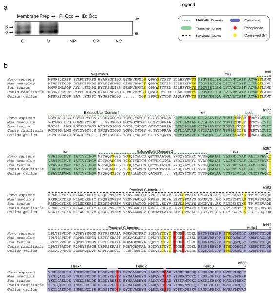
Figure 1.
VEGF-induced occludin phosphorylation sites. (a) Following crude membrane preparation of BREC, occludin was immunoprecipitated, and separated by SDS-PAGE. A portion of the lysate was transferred and probed with rabbit polyclonal anti-Occludin antibody (α-Occ-r) (Zymed) to localize occludin. The remainder of the lysate was separated by SDS-PAGE and three regions of the gel, corresponding to different occludin isoforms, were excised and analyzed by MALDI-TOF MS. C, Control; V, VEGF 15 min; NP, no primary antibody; OP, other primary antibody; NC, no cell (no cell lysate control). (b) Occludin phosphosites identified from VEGF-stimulated endothelial cells. Tryptic peptides of occludin were analyzed by FindMod and data were filtered by inclusion criteria defined in Materials and Methods. Occludin sequence was aligned with the ClustalV algorithm. MS coverage of occludin is indicated by a solid line under the B. taurus sequence. Green shading represents transmembrane domains and blue represents the coiled-coil domain, while other domains are indicated above the aligned residues. Putative phosphosites, red; conserved S/T, yellow.

