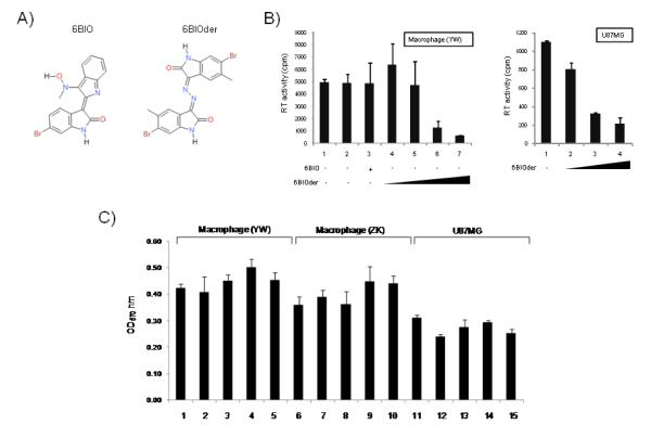
Figure 4. Effect of 6BIO and 6BIOder on HIV-1 replication.
A) Structure of 6BIO: 2-{[[2-(5-bromo-2-oxo-1,2-dihydro-3H-indol-3 ylidene)hydrazino](oxo) acetyl]amino} benzoic acid and 6BIOder: 6-bromo-5-methyl-1H-indole-2,3-dione 3-[(6-bromo-5-methyl-2-oxo-1,2-dihydro-3H-indol-3-ylidene)hydrazone]. B) PMBCs were obtained from the blood of healthy donor (YW), and purified by centrifugation through a layer of Lymphocyte Isolation Medium. Cells were resuspended in serum-free RPMI and plated on culture dish for 1 hour at 37°C. Non-adherent lymphocytes were removed and the adherent monocytes were cultured in RPMI plus 10% heat-inactivated FBS. Macrophages were further differentiated by incubating in 10 ng/ml M-CSF for 1 week with medium change every 2 days. Both macrophages and U87MG cells were infected with 89.6 (MOI:1). 6BIO (lane 3, 10 nM) 6BIOder (lanes 4-7: 0.1, 1, 10, 100 nM) were added to cells six hours after infection. U87MG cells were treated with 6BIOder at 0.1, 1 and 10 nm (left side of panel B). Samples were collected on day 7 for RT assay. Treatments were performed in triplicate and the average plus the standard deviation is displayed. C) MTT assays in two monocyte/macrophage healthy donors and U87MG were performed after treatment with 6BIOder at 10, 100, 1000 and 10,000 nM (lanes 2-5).

