
Figure 7.
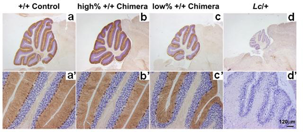
Photomicrographs of cerebellar sections from Lurcher (Lc/+), wildtype (+/+), and Lc/+↔+/+ chimeras taken at both low (top row) and high (bottom row) magnification demonstrating the loss of Purkinje cells in Lc/+ and chimeric mice compared to a control sample. Purkinje cells were stained immunocytochemically with the anti-Calbindin antibody (Chemicon) and appear dark brown in a single monolayer above the blue-stained cerebellar granule cells (cresyl violet counterstain). a Section from a +/+ control cerebellum with normal Purkinje cell number and cerebellar size. b Section from the cerebellum of a high-percentage +/+ chimera with a relatively minor loss of Purkinje cells. c Section from the cerebellum of a low-percentage +/+ chimera showing a relatively major loss of Purkinje cells. d Section from a Lc/+ cerebellum demonstrating the complete loss of Purkinje cells and extreme decrease in cerebellar size. Scale bar = 500 µm a–d, 120 µm a'–d'. Copyright © 2006 by the American Psychological Association. Reproduced with permission from [290]. The use of APA information does not imply endorsement by APA.