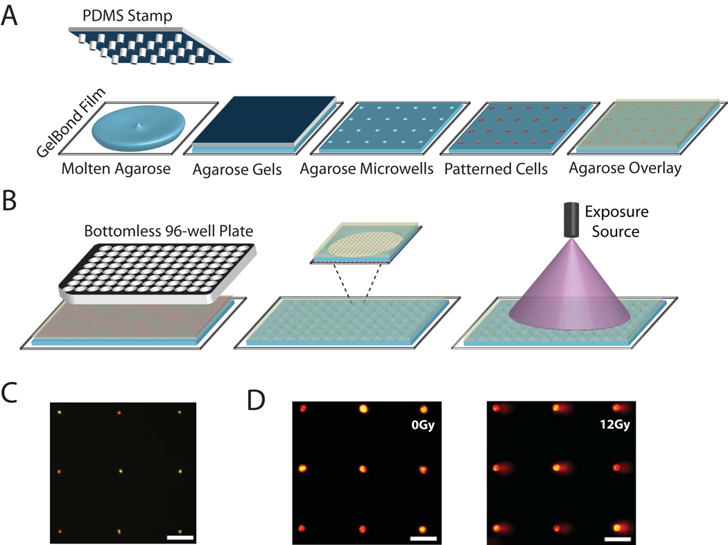
Figure 1.
High throughput comet analysis platform. (A) A microfabricated PDMS stamp with microposts is placed onto molten agarose. After the agarose gels, the stamp is removed and cells are loaded into the microwells by gravity. An agarose overlay encloses cells. (B) To create the CometChip, a bottomless 96-well plate is pressed on agarose gel with embedded cell array that is placed on a glass substrate to create the macrowell platform (each well of the 96 well plate is considered to be a single macrowell). Each macrowell has within it ~300 microwells loaded with cells. (C) Single cell loading in microwells. Two populations of cells stained red and green were loaded concurrently. (D) Arrayed non-irradiated (left) and 12-Gy irradiated (right) microwell comets observed under 10x objective lens. Monohrome images were collected from microscope camera and colored with Red Hot lookup Table using Image J. Horizontal scale bars are 100 µm.

