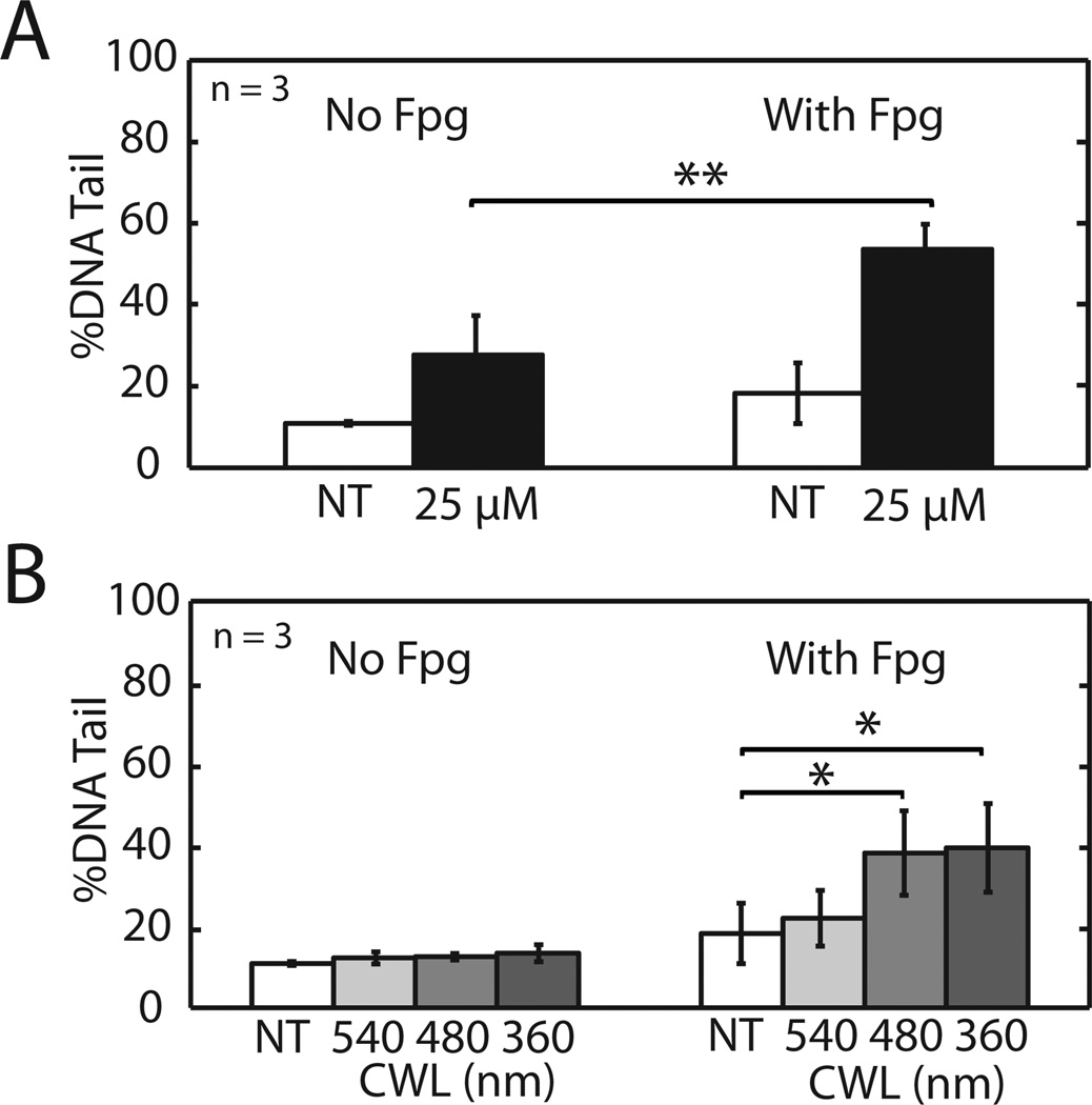
Figure 4.
Oxidative DNA base lesions revealed by the Fpg glycosylase. TK6 cells in the CometChip were exposed to (A) 25 µM H2O2 or (B) 5 min light of different CWLs, lysed and incubated with Fpy glycosylase to reveal oxidative damage. Data and error bars represent averages and standard errors of three independent experiments. Symbols indicate significance according to Student’s T-test:*p < 0.05, **p < 0.01.

