
FIG. 1.
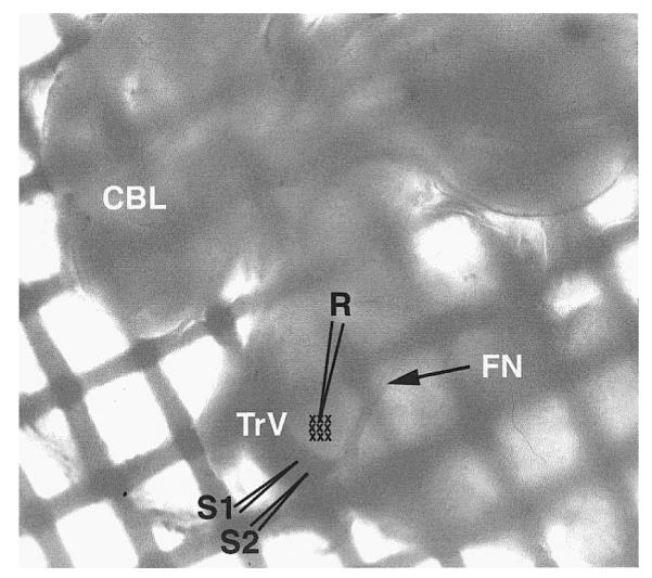
Low-power photomicrograph of a freshly dissected brain stem slice from a postnatal day 6 (PND 6) rat pup within the recording chamber. The trigeminal tract (TrV) and the principal sensory nucleus (PrV) are readily visible and the stimulating (S1 and S2) and recording (R) sites are sketched on the photomicrograph. Barrelette region is also indicated (small crosses). FN, root of the facial nerve; CBL, cerebellum. The slice lies over a nylon mesh grid, mesh size = 500 μm2.