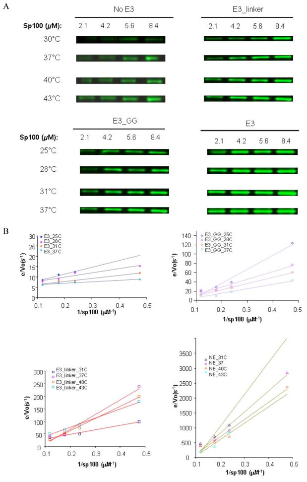
Figure 2.
Temperature dependent enzyme kinetic measurements of the wild-type and mutant E3. (A) Examples of Western blots used to detect the Sp100~SUMO product. The differences in temperatures and reaction times between the E3 variants were necessary to ensure that Sp100~SUMO can be accurately detected. (B) Double reciprocal plots of data shown in (A). The assay mixture (4.2 μL) contained 0.78 μM E1, 0.35 μM E2, 2.6 μM E3, 4 mM ATP, 22.7 μM SUMO, and four Sp100 concentrations: 2.1, 4.2, 5.6, and 8.4 μM. E3 variants used: no E3 (NE), E3_linker, E3_GG insertion, and wild-type E3. Temperatures: 31–43°C, 10 min for NE and E3_linker assays; 25–37°C, 3 min for E3_GG insertion and wild-type E3 assays. Assays were quenched with freshly prepared 360 mM DTT loading buffer (8.5 μL), separated by SDS-PAGE and detected by Western blots using an anti-SUMO-1 antibody.

