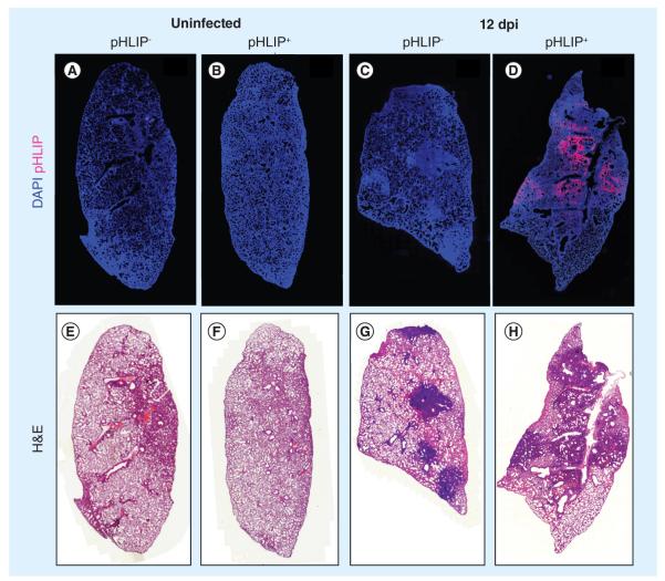
Figure 2. pH (low) insertion peptide targets infected but not uninfected mouse lungs.
Representative whole-lung images from three independent experiments (n ≥4). pHLIP was administered to influenza A/Puerto Rico/8/34-infected mice on day 10 postinfection. (A–D) DAPI (blue) and pHLIP (red). (B & D) At 12 dpi, pHLIP can only be detected in infected lungs.
(A & C) Control lungs from uninfected and infected mice. (E–H) H&E staining of the same samples. Darkened areas indicate regions of immune cell infiltration and inflammation.
dpi: Days postinfection; H&E: Hematoxylin and eosin; pHLIP: pH (low) insertion peptide.

