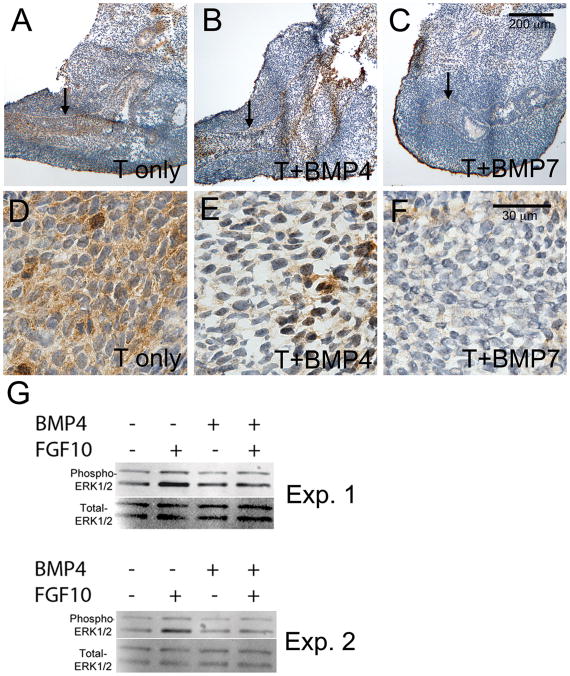
Figure 7. FGF10-induced ERK1/2 activation is reduced in BMP treated UGS tissues.
E15 UGS tissues were cultured for 48 hours in the presence of testosterone only (A, D), testosterone + BMP4 (B, E), or testosterone + BMP7 (C, F). At the end of the culture period, tissues were treated with FGF10 for 20 minutes and then processed for analysis of ERK1/2 activation. Tissue sections were evaluated using immunostaining with an antibody to detect phosphorylated (activated) ERK1/2. Significant staining (brown stain) was present in the testosterone only treatment and reduced in the UGE following BMP4 or BMP7 treatments (UGE indicted by the arrows in A-C and shown at higher magnification in D-F). Overall reductions in UGS phospho-ERK1/2 were also detected by Western blotting following BMP4 treatment in four independent experiments (G and data not shown). Results from two independent experiments (Exp. 1 and Exp. 2) are shown in G. Tissue sections were counterstained with hematoxylin (purple stain in A-F). Scale bars for A-C and D-F are shown in panels C and F respectively.

