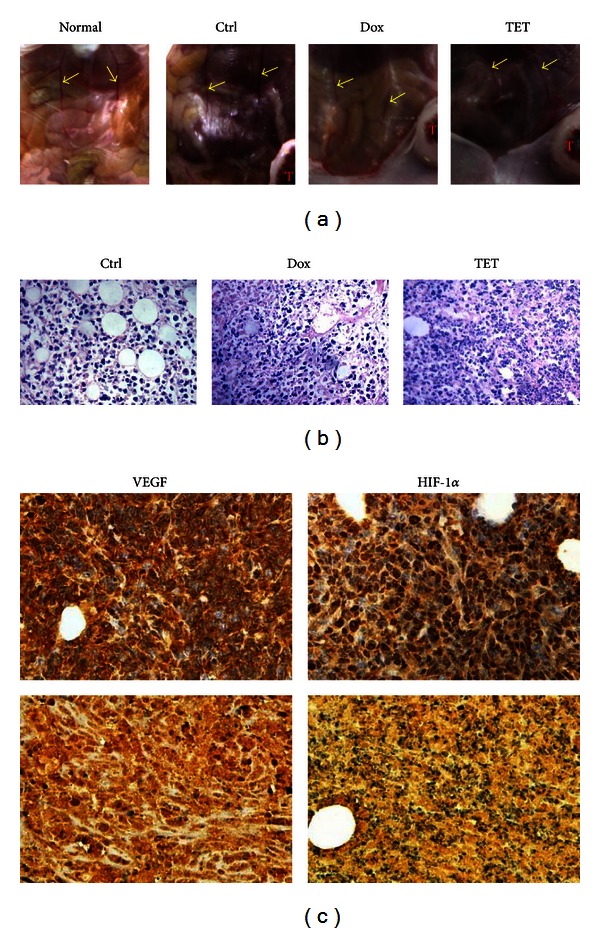
Figure 5.

TET suppressed tumor angiogenesis in 4T1 tumor bearing mice. (a) The blood vessel diameter of TET treated tumor bearing mice. Picture shows the diameter of blood vessel on mice abdomen for each mouse. (b) Hematoxylin & Eosin staining of tumor tissues. Retrieved tumor samples were fixed, embedded, and subjected to H&E staining. Representative images are shown (magnification, ×400). (c) VEGF and HIF-1α expressions in tumor tissues. Representative staining results are shown: 4T1 tumor-bearing BALB/c mice (top panel) and TET treated mice (bottom panel) (magnification, ×400).
